Dental Fluorosis
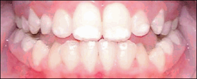
Spots on your teeth? You might have dental fluorosis.
(Video running time: 54 seconds)
Overdosed on Fluoride: The Dental Fluorosis Problem
(Video running time 4:02 minutes)
Many thanks to FAN TV for these informative videos.
Even fluoridation advocates agree that ingestion of fluoridated water when teeth are forming can cause dental fluorosis.
The traditional “Dean Index” lists four stages of visible damage to the tooth enamel ranging from mild dental fluorosis (faint opaque white marks on up to 50% of the tooth’s surface), mild, moderate, and severe dental fluorosis (the whole surface of the tooth is affected, dark brown discoloration, sometimes with pitting and flaking ) .
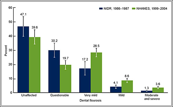
SOURCES: CDC/NCHS, National Health and Nutrition Examination Survey (NHANES), 1999-2004 and National Institute of Dental Research, National Survey of Oral Health in U.S. School Children, 1986-1987.
THE TOOTH SURFACE INDEX OF FLUOROSIS (TSIF SCALE) IDENTIFIES SEVEN STAGES:
Score = 0: Enamel shows no evidence of fluorosis.
Score = 1: Enamel shows definite evidence of fluorosis, namely areas with parchment-white color that total less than one-third of the visible enamel surface. This category includes fluorosis confined only to incisal edges of anterior teeth and cusp tips of posterior teeth (“snowcapping”).
Score = 2: Parchment-white fluorosis totals at least one-third of the visible surface, but less than two-thirds.
Score = 3: Parchment-white fluorosis totals at least two-thirds of the visible surface.
Score = 4: Enamel shows staining in conjunction with any of the preceding levels of fluorosis. Staining is defined as an area of definite discoloration that may range from light to very dark brown.
Score = 5: Discrete pitting of the enamel exists, unaccompanied by evidence of staining of intact enamel. A pit is defined as a definite physical defect in the enamel surface with a rough floor that is surrounded by a wall of intact enamel. The pitted area is usually stained or differs in color from the surrounding enamel.
Score = 6: Both discrete pitting and staining of the intact enamel exist.
Score = 7: Confluent pitting of the enamel surface exists. Large areas of enamel may be missing and the anatomy of the tooth may be altered. Dark-brown stain is usually present.
SOURCE: Horowitz HS, et al. (1984). A new method for assessing the prevalence of dental fluorosis–the Tooth Surface Index of Fluorosis. Journal of the American Dental Association 109(1):37-41.
![FIGURE 1: Martinez-Mier EA, Soto-Rojas AE. (2010). Differences in exposure and biological markers of fluoride among White and African American children. Journal of Public Health Dentistry 70:234-40.“Of the 62.5 percent of the White children [from Indianapolis, Indiana] who presented with dental fluorosis upon examination, 41.3 percent had a maximum score of 1 and only 21.2 percent of the children had a maximum score of 2. Of the 80.1 percent of African American children who had dental fluorosis, a maximum score of 1 was assigned to 50.5 percent of the children, 15.4 percent were assigned a maximum score of 2, 1.5 percent had a maximum score of 3, and 12.7 percent were assigned the highest score of 5. Differences in severity were also statistically significant (P < 0.001).” SOURCE: Martinez-Mier EA, Soto-Rojas AE. (2010). Differences in exposure and biological markers of fluoride among White and African American children. Journal of Public Health Dentistry 70:234-40.](http://www.cleanwatersonomamarin.org/wp-content/uploads/2016/01/graph_martinez-2010_fluorosis-and-race_fr_502.jpg)
| Dental Fluorosis Rates in Augusta & Richmond County, Georgia |
|||
| Residence/Race | No Fluorosis (TSIF Score = 0) |
Very Mild/Mild Fluorosis (TSIF Score = 1 – 3) |
Moderate/Severe Fluorosis (TSIF Score = 4 – 7) |
| City/Black | 19.6% | 63.7% | 16.7% |
| City/White | 18.2% | 72.7% | 9.1% |
| County/Black | 47.8% | 48.9% | 3.3% |
| County/White | 44.9% | 55.1% | 0% |
| SOURCE: Williams JE, Zwemer JD. (1990). Community water fluoride levels, preschool dietary patterns, and the occurrence of fluoride enamel opacities. Journal of Public Health Dentistry 50:276-81. | |||
Dental fluorosis as a marker for fluoride neurotoxicity: findings from a study by Anna L.Choi et al (2014)
“Common sense should tell us that if a poison circulating in a child’s body can damage the tooth-forming cells, then other harm also is likely.” – Dr. John Colquhoun (1997).
Professor Howard Pollick. Testimony that, to reduce the possibility of dental fluorosis, young babies should not ingest infant formula prepared with fluoridated water. Informational presentation on proposed water fluoridation before the City of San Diego Joint Rules Committee & Special City Council Meeting (July 18, 2007). Chair Scott Peters. Running time 02:07 minutes.